Coronavirus disease 2019 (COVID-19) is
defined as illness caused by a novel coronavirus. It was initially reported in
Wuhan City, China in December 2019 and WHO declared COVID-19 as a pandemic in
March 2020.
COVID-19 suspected patients may present with
uncomplicated (mild) illness as well as with severe disease. As per the initial
reports, 81% of patients have mild (absent or mild pneumonia), 14% have severe
(hypoxia, dyspnoea, >50% lung involvement within 24-48 hours), 5% have
critical (shock, respiratory failure, multiorgan dysfunction), and 2.3% have
fatal disease. Patient with severe, critical and fatal disease is the people
who need airway management, especially patients having type 1 respiratory
failure, ARDS, septic shock or MODS.
Rapid sequence intubation is the method
of choice for securing airway in the Emergency Department. While managing airway
of patient with COVID-19, in addition to the conventional 7 P’s of RSI, an
eighth ‘P’ should be considered i.e. Personal protection. Personal protection
includes the provider safety which should be given utmost care and considering
this, the nomenclature RSI (Rapid sequence intubation) may be renamed as
protected intubation. The 8 P’s of protected intubation is given in Table 1
1
|
Personal
Protection
|
2
|
Preparation
|
3
|
Preoxygenation
|
4
|
Pre-treatment
|
5
|
Paralysis
and sedation
|
6
|
Position
|
7
|
Placement
of tube
|
8
|
Post
Intubation care
|
Table 1: 8 P’s of Protected Intubation
There is no high-level evidence for
these modifications – at best the evidence is Level C – consensus/expert
opinion
1. Personal
Protection
Protection of team is
most important while taking care of COVID19 patient, hence the patient should
be provided a 3-ply surgical mask (Fig 1) so as to minimise droplet spread as
soon as he enters the health care facility. |
| Figure 1: Patient with 3-ply surgical mask |
All persons taking care of the patient should wear Level 3 PPE is to be worn for any AGP. List of AGP is given in the table below (Table 2). It is advisable to wear 3 pairs of gloves and the outermost glove should be removed soon after intubation so as to minimise soiling of other areas. To prevent shield/ fogging, it would be better to cover the inner side of shield/ goggles with a layer of transparent hand sanitizer.
Table 2: Aerosol Generating Procedures (AGP)
|
|
Cardiopulmonary
resuscitation
|
Bag valve mask
ventilation
|
Non-invasive
positive pressure ventilation
|
High flow
nasal cannula
|
Intubation and
Extubation
|
Nebulisation
|
Bronchoscopy
|
Tracheostomy
|
Suctioning
|
Cricothyroidotomy
|
Oro/
Nasopharyngeal swab collection
|
Dental
procedures involving high speed drill
|
Minimum number of personnel’s (3 person) inside the room and a person should be just outside the room as a runner (In level 3 PPE) who will work as an assistant if needed.
Anything entering within 2 meter radius of patient should be considered as contaminated (Fig 2).
 |
| Figure 2: Area of contamination around the patient |
 |
| Figure 3: Droplet spread |
 |
| Figure 4: Aerosol spread |
2. Preparation
It is ideal to
have a pre-prepared COVID intubation tray (Fig: 5, 6,7,8) which will have
all
the equipment’s including equipment’s for failed airway. It is ideal to have
two
functional IV access (preferably in proximal large vein). Meticulous airway
assessment
and plan for failed airway should be done during the preparation phase
and all the team
members should be well aware of the plan for intubation. Rough
layout of room for
airway management is depicted in figure 9.
 |
| Figure 7: Stylet, gum
elastic bougie, supraglottic airway device, oropharyngeal airway and McCoy
laryngoscope blade as rescue devices in failed airway plan. |
 |
| Figure 8: eFONA set – comprising
of No: 11 scalpel blade, gauze pack, Cuffed 5.5 or 6 size ET tube and
tracheostomy tube |
 |
| Figure 9: Room layout |
3. Preoxygenation
Passive
preoxygenation should be done for minimum of 5 minutes. Position the patient
for preoxygenation by keeping the head elevated by 25-45 degree. Separate
oxygen
source for preoxygenation with Non rebreather mask (NRBM) kept at a flow
rate of
10-15L/min (Fig 12) and for apnoeic oxygenation with nasal cannula kept
at flow rate
of 4-6L/min (Fig 13) (NODESAT – Nasal oxygen during efforts
securing a tube).
NRBM to be kept over the 3-ply mask to minimise
aerosolization (Fig 10). Lowest
possible gas flow should be used to maintain
oxygen saturation above 92%. VAPOX
(Ventilator Assisted PreOXygenation) should
be avoided as far as possible and if at all
used, a non-vented NIV mask should
be used and filters should be incorporated in the
circuit. Noncompliance of
patient for preoxygenation may be mitigated by the use of
dissociate dose of
Ketamine (0.5-1mg/kg).
Bag valve mask
device with viral filter can be used for preoxygenation but positive
pressure
ventilation should be avoided as far as possible. Instead of E-C clamp
technique, a 2 hand Vice grip (V-E grip) gives a better seal and is the
preferred method
(Fig 14, 15). A better seal is obtained by using NIV mask (non-vented)
strapped to the
patient (Fig 16). This
should also have a filter proximal to the mask.
Oxygen source
should be turned off before removing the mask so as to ensure provider
safety.
 |
| Figure 10: 3-ply surgical mask over which NRBM is applied |
 |
| Figure 11: Graph showing aerosol
dispersion while using various modalities of treatment |
 |
| Figure 12: Nasal prongs for apnoeic oxygenation (3-ply surgical mask not shown in the picture) |
 |
| Figure 13: NRBM for preoxygenation along with nasal prongs (3-ply surgical mask not shown in the picture) |
 |
| Figure 14: Double E-C Clamp technique giving head tilt chin lift |
 |
| Figure 15: V-E technique giving jaw thrust |
 |
| Figure 16: Bag valve mask with reservoir bag fitted with a NIV mask (non-vented) and filter proximal to the mask |
4. Pre-treatment
Antimuscarinic
agent Glycopyrrolate 0.2mg IV may be used to decrease secretions. Lidocaine 1.5
– 2
mg/kg IV to be used to decrease the incidence of coughing during intubation.
When bronchospasm is
a concern, it is prudent to use metered dose inhaler
rather than nebuliser in order to decrease the risk
of aerosolization.
If time permits,
adequate hemodynamic resuscitation should be the norm and consider
fluid-sparing
approach due to concerns for third-spacing. Prior to induction, consider
having vasopressors
prepared/ running (norepinephrine 5-35 mcg/min) –
particularly for patients with hypotension, signs
of impaired perfusion, or
elevated shock index (HR÷SBP > 1) or keep push dose pressor handy and
use it
as a bridging agent for buying time to initiate pressor infusion if patient
becomes
haemodynamically unstable.
5. Paralysis
and induction
Ketamine 1– 2mg/kg
of ideal body weight or Etomidate 0.15 – 0.3mg/kg of total body weight are the
preferred induction agents. Lower doses of induction agents (aliquots of
0.5mg/kg of Ketamine until
sedation is achieved) are ideal in haemodynamically
unstable patients so as to prevent post intubation
adverse events.
Succinylcholine
1.5 – 2mg/kg of total body weight or Rocuronium 1.2 – 1.6mg/kg total body
weight
are the preferred paralytic agents of choice. Rocuronium has an added
advantage of longer safe
apnoea time compared to Succinylcholine. Higher dose
of muscle relaxant to be considered in
haemodynamically unstable patient.
Complete paralysis should be ensured before intubating so as to
prevent
coughing.
6. Position
Sniffing
position is the conventional position for intubation but variations may be
tried as per the skill
of the provider. Bed Up Head Elevated (BUHE) position is
one such modification which can be done
which gives an added advantage of
better oxygenation and longer safe apnoea time (Fig 17, 18, 19
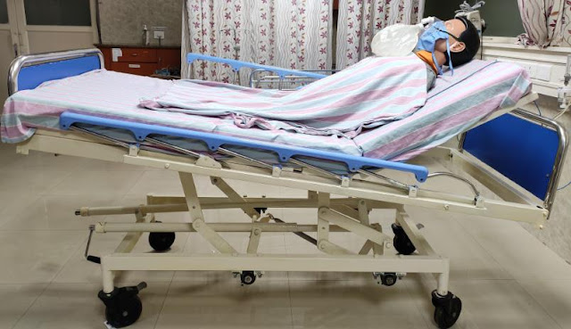
20).
 |
| Figure 17: Bed kept flat |
 |
| Figure 18: Foot end elevated (Trendelenburg position) |
 |
| Figure 19: Head end elevated to 30 degree |
 |
| Figure 20: Head roll under the neck |
7. Placement
of tube
Intubation to be done by the most
experienced person in the team and preferably with a video laryngoscope so as
to increase the distance between the provider and the patient. Use barrier
enclosure if available or transparent polythene sheet so as to contain droplet
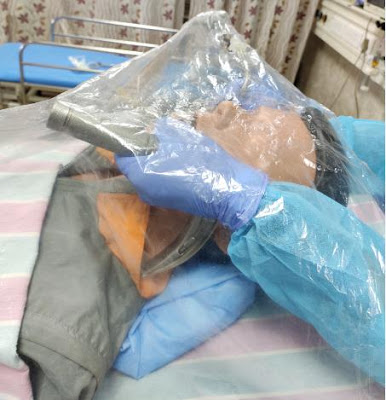
spread. Intubation should be done beneath the polythene sheet (Fig 21). Proximal
end of the ET tube should be plugged with gauze so as to minimise contamination
(Fig 22). If bougie is preloaded then also the proximal end of tube should be
plugged with gauze (Fig 23). Kiwi grip may be tried so that the airway
assistant need not come in close proximity to the patient (Fig 24). Tube is to
be fixed at a premeasured length. Visualisation of black line passing through
the cords so as to make sure it’s endotracheal. Confirming the depth of the
endotracheal tube is extremely difficult using auscultation while wearing
isolation suits. It is recommended instead to observe bilateral chest
expansion, ventilator breathing waveform, and respiratory parameters. End-tidal
CO2 is a better indicator of successful tracheal intubation, as oxygen
saturation is not always increased immediately after intubation in these
patients, because the oxygen exchange is significantly impaired. Bedside
ultrasonography is another modality which can be used for confirmation of ET
tube position. Cuff is to be inflated before initiating ventilation and cuff
pressure to be monitored so as to minimise leak. Remove outer glove soon after
intubation to limit contamination.
 |
| Figure 21: Transparent polythene sheet beneath which intubation is being done so as to minimise droplet spread |
 |
| Figure 22: Prechecked ET tube preloaded with malleable stylet and proximal end plugged with gauze |
 |
| Figure 23: ET tube preloaded with gum elastic bougie and proximal end plugged with gauze |
 |
| Figure 24: Kiwi grip so as to minimise the number of personnel coming in close proximity to the patient |
8. Post
intubation care
Analgesia first
followed by sedation so as to prevent patient machine asynchrony.
Opioid as
analgesic and benzodiazepine as a sedative agent as per the haemodynamic
profile of the patient will be ideal. Avoid unnecessary ventilation
disconnection. If
disconnection is needed, put PPE, put ventilator on standby
and clamp ET tube.
Disconnection of circuit should be done distal to the
filter. Use closed suction
whenever possible. Ventilatory parameters should be
pre-set should be a part of the
preparation phase (Fig 25). Both the limbs of
circuit should have filters. Initial
ventilator settings include Volume assist
control mode with tidal volume of 6cc/kg
IBW, Inspiratory flow to be kept
between 60-80L/min respiratory rate of 16-18 breaths
per minute, FiO2 100%,
PEEP of 5cm H2O which should be titrated as per ARDSnet
protocol. Saturation
should be maintained above 96% and plateau pressure to be
maintained below
30cm H2O.
Nasogastric tube
to be inserted after patient is being ventilated safely. After the
procedure,
discard the disposable items properly and proper disinfection of reusable
items
should be done. Doffing should also be done in a proper manner.
 |
| Figure 25: Ventilator pre-set
and filters in both inspiratory and expiratory limb of circuit |
Failed Airway
In the event of a
failed intubation attempt [Cannot Intubate Cannot Ventilate (CICO)], a second
generation supraglottic airway device (SGD) should be used as a temporary bridging method. Proximal end of
SGD should be plugged with gauze so as to minimize contamination (Fig: 26).
Ventilation should be commenced only after ensuring that appropriate sized SGD
is fixed at adequate depth, and the cuff is fully inflated. |
| Figure 26: Supraglottic airway device with proximal end plugged with gauze |
Emergency Front of Neck Access (eFONA) –
scalpel bougie technique to be considered early in the course of failed airway
Cardiopulmonary resuscitation
Prearrest
If at risk of cardiac arrest, consider
shifting the patient to negative pressure room proactively to minimize exposure.
Close the door to prevent adjacent area contamination if negative pressure room
is unavailable.
Considerations before initiating
cardiopulmonary resuscitation
1 Is
it too late?
Mortality from cardiac arrest secondary
to COVID19 is approximately 13% which is caused by hypoxaemia and/or
myocarditis. Reconsider resuscitation if in end stage disease state.
2 What
about Do Not Resuscitate Status?
Consider DNR status before attempting
resuscitation as the severity of illness is more in elderly age group as well
as with comorbid illness.
3 What
about the adequacy of Resources?
In a pandemic where resources are
outnumbered by needs, think before initiating a full-fledged resuscitation. When
considering personnel’s and ventilators as a scarce resource, a prevented resuscitation
can result in better resource allocation.
Considerations during
cardiopulmonary resuscitation
3 persons should suffice for performing
cardiopulmonary resuscitation. Level 3 PPE (Disposable gown, gloves, FFP3
respirator and eye protection) should be worn by all team members especially when
performing chest compression, Bag valve mask ventilation and intubation. So as
to reduce the number of personnel’s, mechanical chest compression devices
should be considered during resuscitation.
Intubation attempts should be minimized
so the most experienced person in airway management should take care of the
victim’s airway. Chest compressions should be paused during attempted
intubation to improve the first pass success. Video laryngoscope should be the
choice of equipment for intubation. After intubation, minimise disconnections
of circuit so as to reduce aerosolization.
In instances where intubation is
delayed, bag valve mask or supraglottic airway with a filter should be used for
ventilation. A tight seal should be obtained during bag valve mask ventilation
to reduce aerosolization.
Filter should be attached to a bag valve
mask, supraglottic airway or before connecting to mechanical ventilator to
reduce aerosolization.
If already intubated at the time of
cardiac arrest, don’t try to disconnect the patient from ventilator so as to
maintain closed circuit. Ventilator settings such as FiO2, mode of ventilation,
trigger, respiratory rate and alarms should be readjusted.
Considerations after cardiopulmonary
resuscitation
While considering post cardiac arrest
care, the conventional ABCDE approach should be applied. Apart from the
conventional management things which should be considered are:
1 1. Optimise
ventilatory settings according to the patient’s clinical condition.
2 2. Local
infection control practises after resuscitation for doffing and patient
transport.
About the author: Dr. Jebu A Thomas (MBBS, DNB(EM), MRCEM, MNAMS) currently works as Assistant Professor in Emergency Medicine at Pushpagiri Institute of Medical Sciences, Thiruvalla, Kerala. He can be reached at dr.jebu@gmail.com
About the author: Dr. Jebu A Thomas (MBBS, DNB(EM), MRCEM, MNAMS) currently works as Assistant Professor in Emergency Medicine at Pushpagiri Institute of Medical Sciences, Thiruvalla, Kerala. He can be reached at dr.jebu@gmail.com


Comments
Post a Comment